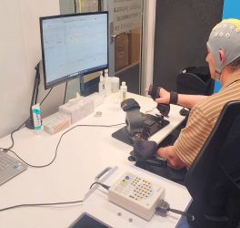
Multiple sclerosis (MS) is a neurological disease that usually leads to permanent disabilities. It affects around 2.9 million people worldwide, and around 15,000 in Switzerland alone. One key feature of the disease is that it causes the patient’s own immune system to attack and destroy the myelin sheaths in the central nervous system. These protective sheaths insulate the nerve fibres, much like the plastic coating around a copper wire. Myelin sheaths ensure that electrical impulses travel quickly and efficiently from nerve cell to nerve cell. If they are damaged or become thinner, this can lead to irreversible visual, speech and coordination disorders.
So far, however, it hasn’t been possible to visualise the myelin sheaths well enough to use this information for the diagnosis and monitoring of MS. Now researchers at ETH Zurich and University of Zurich, led by Markus Weiger and Emily Baadsvik from the Institute for Biomedical Engineering, have developed a new magnetic resonance imaging (MRI) procedure that maps the condition of the myelin sheaths more accurately than was previously possible. The researchers successfully tested the procedure on healthy people for the first time.
In the future, the MRI system with its special head scanner could help doctors to recognise MS at an early stage and better monitor the progression of the disease. The technology could also facilitate the development of new drugs for MS. But it doesn’t end there: the new MRI method could also be used by researchers to better visualise other solid tissue types such as connective tissue, tendons and ligaments.
Quantitative myelin maps
Conventional MRI devices capture only inaccurate, indirect images of the myelin sheaths. That’s because most of these devices work by reacting to water molecules in the body that have been stimulated by radio waves in a strong magnetic field. But the myelin sheaths, which wrap around the nerve fibres in several layers, consist mainly of fatty tissue and proteins. That said, there is some water – known as myelin water – trapped between these layers. Standard MRIs build their images primarily using the signals of the hydrogen atoms in this myelin water, rather than imaging the myelin sheaths directly.


