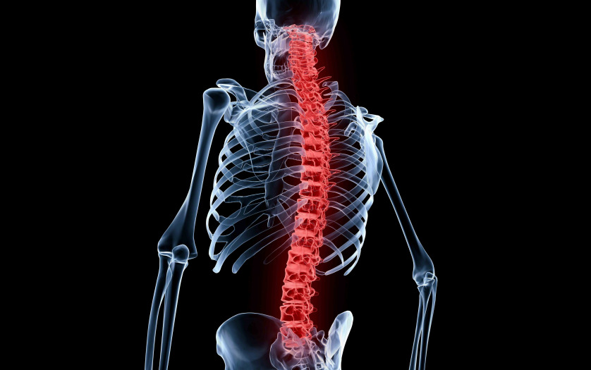
A subgroup of patients recovering from severe spinal-cord injury can develop an autoimmune response directed against spinal cord proteins that may associate with pain development according to a new study by researchers at The Ohio State University Wexner Medical Center and collaborators in Germany, Austria, Italy, Canada and Switzerland.
The researchers found that, after spinal-cord injury, the immune system of some patients produces antibodies that bind to the spinal cord and living neurons. The researchers state that such autoantibodies could lead to the development pain or even interfere with the response to rehabilitation. The findings could lead to new treatments for spinal-cord injury patients who have certain antibody profiles.
The researchers published their findings in the journal Neurology: Neuroimmunology & Neuroinflammation, a journal published by the American Academy of Neurology (AAN).
“To our knowledge, this is the first study to demonstrate that, after spinal-cord injury, patients may increasingly develop autoantibodies that bind to the spinal cord and to the surface of neurons,” said corresponding author Dr. Jan Schwab, the William E. Hunt & Charlotte M. Curtis Chair and a professor of neurology and neurosciences at Ohio State.
“As such, our findings indicate that antibody-mediated autoimmune responses may emerge in some patients about three weeks after spinal-cord injury. So far, we know about antibody-mediated autoimmune syndromes developing in patients with cancer, but that it can also occur in patients after spinal cord injury is striking” said Schwab, who is also medical director of the Belford Center for Spinal Cord Injury and a Scholar of the Chronic Brain Injury Initiative at Ohio State.
The study by Schwab and Dr. Romana Hoeftberger, a Professor of Neuropathology at the Medical University in Vienna (Austria), applied and extended diagnostic methods which were earlier established to detect autoimmune diseases of the brain, such as autoimmune encephalitis. “Here, the ability to become able to diagnose patients with autoimmune encephalitis was crucial. After reaching a diagnosis, these patients can now be successfully treated, which was not possible 15 years ago. In analogy, extending this clinically validated toolbox enabled us to reveal novel autoimmune signatures after spinal cord injury, as first and necessary step,” Schwab noted.
How the study was done
Schwab and his collaborators compared blood-serum antibodies from patients with severe (motor complete and motor incomplete) spinal cord injuries and from control patients over time. The capacity of antibodies to recognize and bind to spinal cord tissue proteins as well as living dorsal root ganglia neurons was tested in a blinded manner.
In addition, the researchers examined archival human spinal tissue samples at time points ranging from the time of injury to several months later. They looked for B- and Plasma cells as cellular origin of the antibody synthesis at the lesion site in spinal-cord injury compared to unaltered control cord tissue.
Key findings included:
- Robust and emerging autoantibody binding was restricted to only a subpopulation (16%) of spinal-cord injury patients; it was absent in controls.
- Autoantibody binding was detected mainly in a specific region of the spinal cord involved in sensory-motor integration (i.e., the substantia gelatinosa).
- Autoantibody binding was associated with patients who required neuropathic pain medication.
- The study of archival spinal-tissue lesions showed B-cell infiltration in 27% of the spinal-cord injury patients, the presence of plasma cells in 9%, synthesizing both IgG and IgM antibodies in areas of activated complement deposition. Antibody synthesis in an area of complement deposition if self-sufficient mechanism to propagate cell death.
- Analysis of cerebral spinal fluid from an additional single patient demonstrated IgM antibody synthesis with late re-opening of the blood spinal cord barrier.
“Overall, our study revealed a new and unexpected autoimmune signature that affects a subpopulation of patients after spinal-cord injury and qualifies as modifiable, potential mechanism to reduce pain and improve quality of life in spinal cord injured patients,” Schwab said.